Answer:
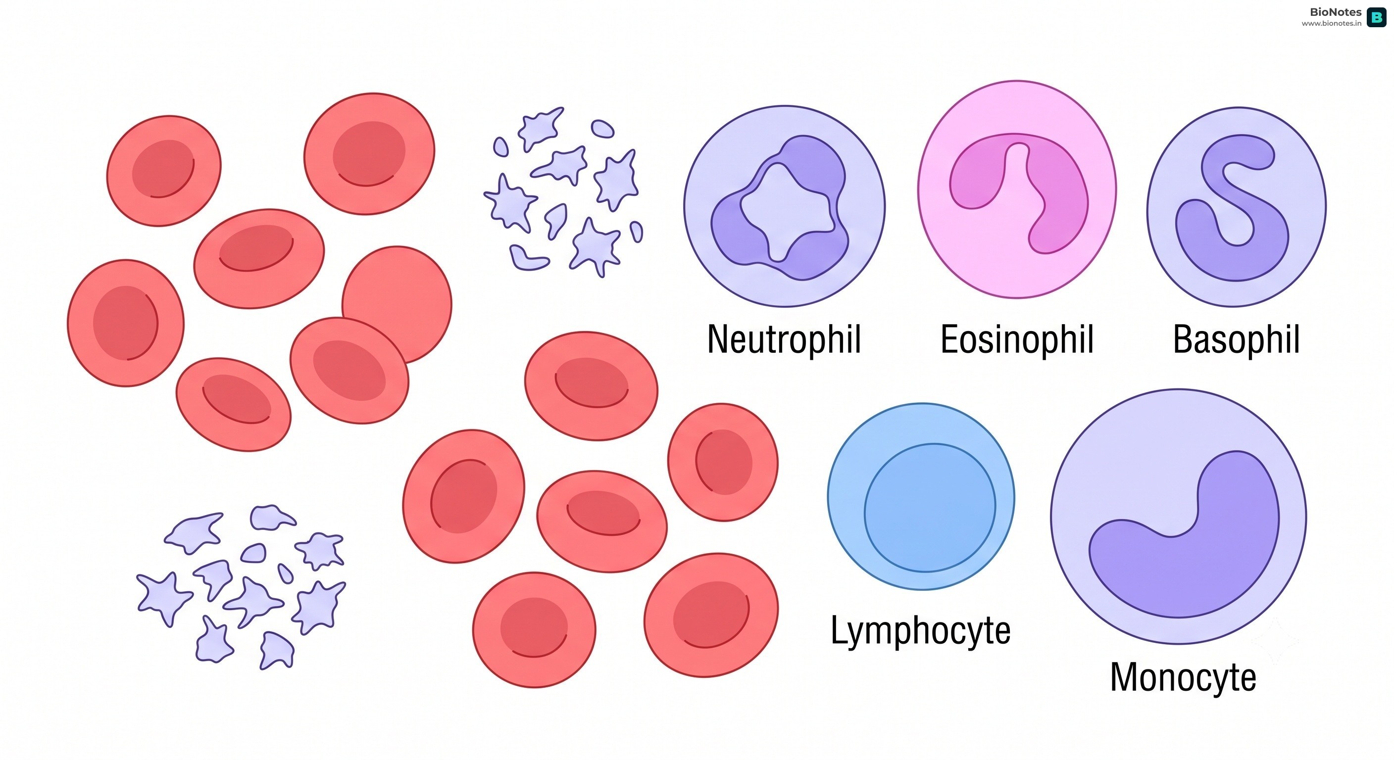
- Erythrocytes (RBCs): Transport respiratory gases (oxygen and carbon dioxide) using haemoglobin.
- Leucocytes (WBCs): Part of the immune system; defend the body against infections.
- Platelets (Thrombocytes): Involved in the coagulation or clotting of blood to prevent excessive blood loss.
Answer:
- Fibrinogen: Essential for blood clotting (coagulation).
- Globulins: Primarily involved in the body's defense mechanism (immunity).
- Albumins: Help in maintaining the osmotic balance and pressure of the blood.
Answer:
(a) Eosinophils - (iii) Resist Infections
(b) RBC - (v) Gas transport
(c) AB Group - (ii) Universal Recipient
(d) Platelets - (i) Coagulation
(e) Systole - (iv) Contraction of Heart
Answer:
Blood is considered a connective tissue because it originates from the mesoderm (like other connective tissues) and it connects different parts of the body by transporting nutrients, gases, and hormones, thus integrating the functions of various organs.
Answer:
- Blood: Contains RBCs, WBCs, and platelets; rich in proteins; carries oxygen and nutrients to tissues; red in color.
- Lymph: A colorless fluid; contains specialized lymphocytes; has fewer proteins and no RBCs; acts as a middleman between blood and tissues, especially for fat absorption.
Answer:
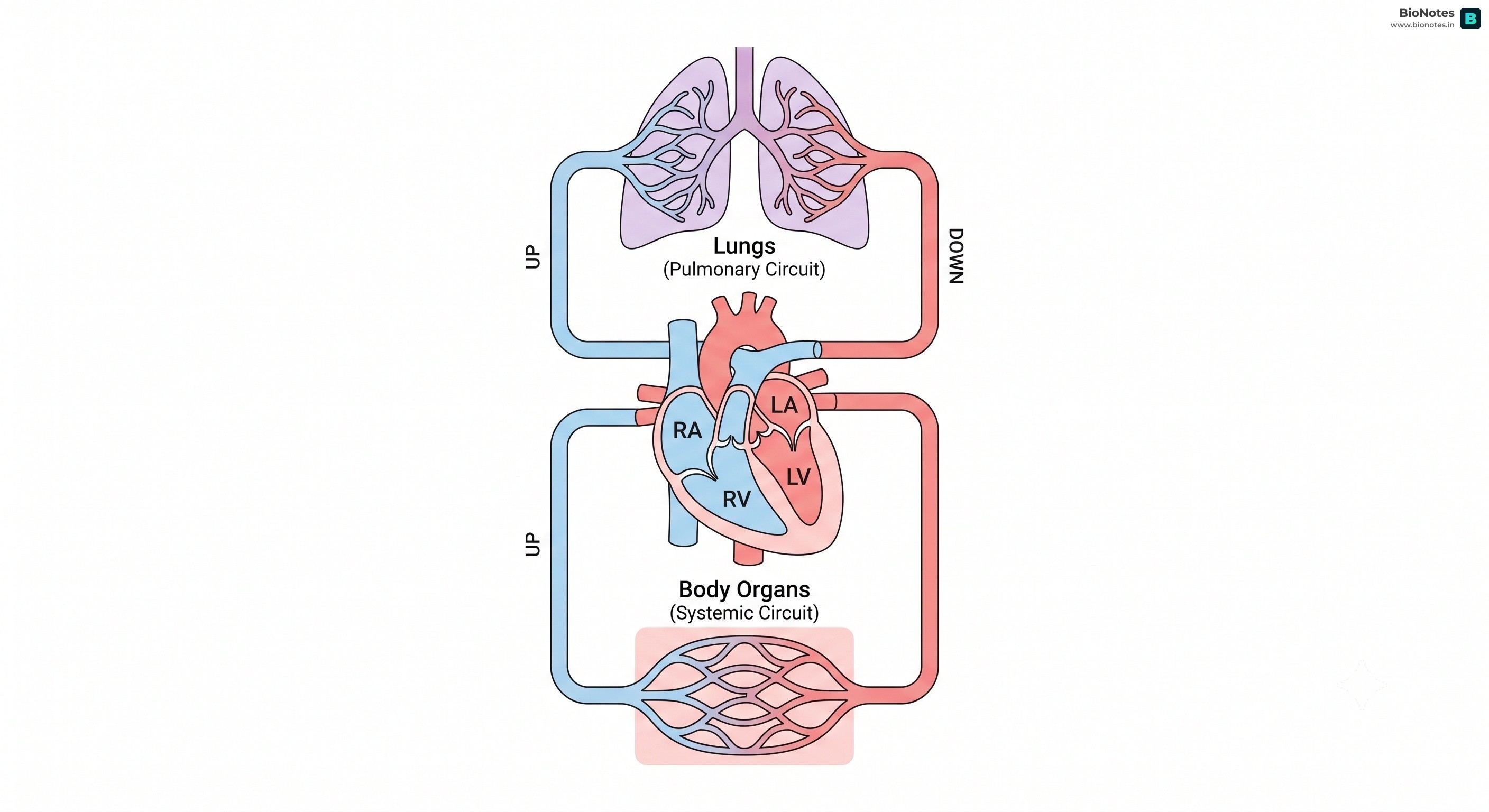
Double Circulation means that the blood passes through the heart twice during one complete cycle through the body. It consists of:
- Pulmonary Circulation: Between the heart and the lungs.
- Systemic Circulation: Between the heart and the rest of the body tissues.
Significance: It prevents the mixing of oxygenated and deoxygenated blood, ensuring an efficient supply of oxygen to all body parts, which is crucial for the high metabolic demands of mammals and birds.
(a) Blood and Lymph
(b) Open and Closed system of circulation
(c) Systole and Diastole
(d) P-wave and T-wave
Answer:
(a) Blood vs. Lymph: Blood has RBCs and is red; Lymph lacks RBCs and is colorless.
(b) Open vs. Closed: In an open system (arthropods), blood flows into body cavities (sinuses). In a closed system (chordates), blood is always confined within blood vessels.
(c) Systole vs. Diastole: Systole is the phase of contraction of the heart chambers. Diastole is the phase of relaxation.
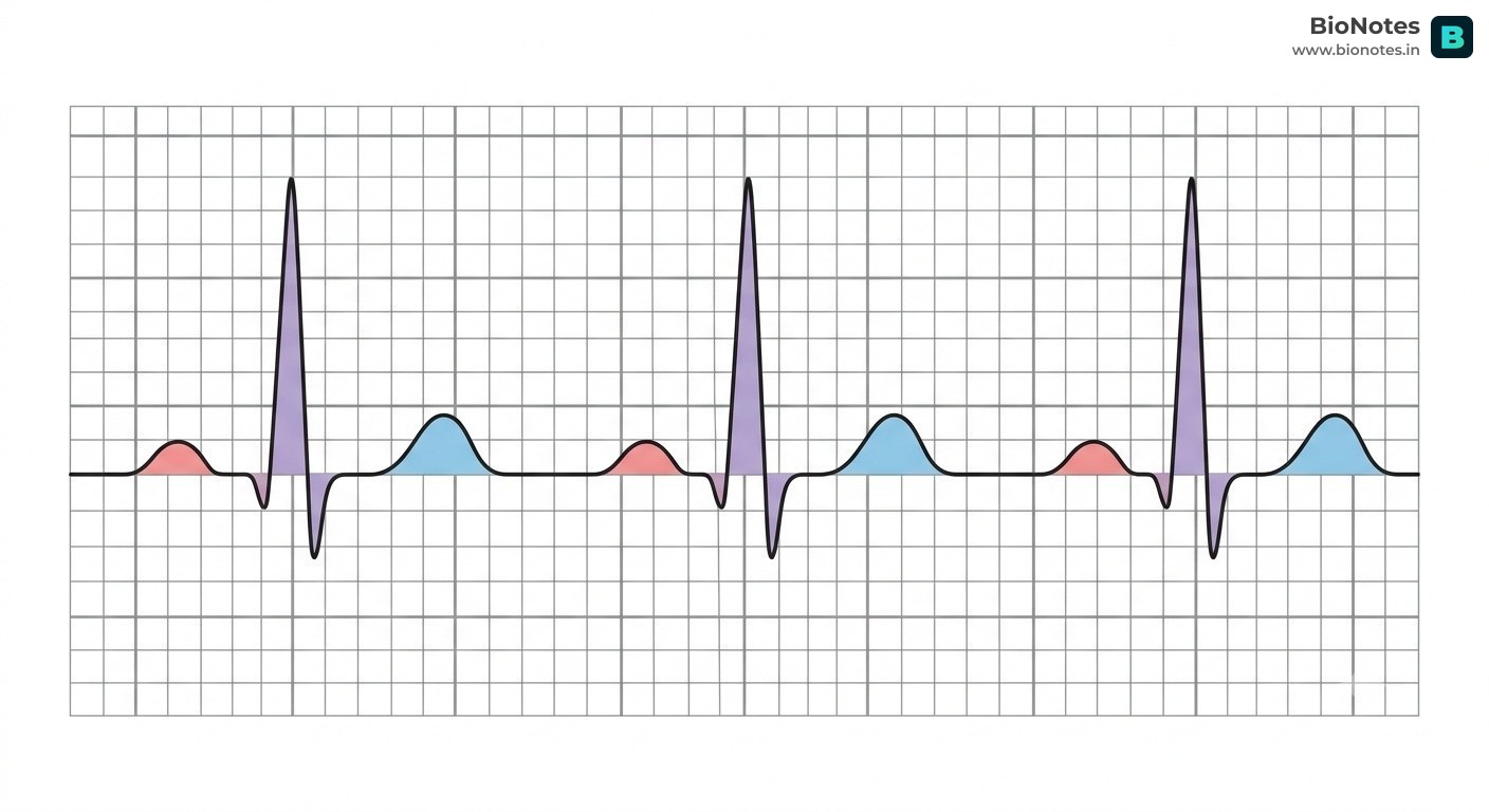
(d) P-wave vs. T-wave: The P-wave represents the depolarization (excitation) of the atria. The T-wave represents the repolarization (return to normal) of the ventricles.
Answer:
- Fishes: 2-chambered (one atrium, one ventricle). Single circulation.
- Amphibians/Reptiles: 3-chambered (two atria, one ventricle). Incomplete double circulation (blood mixes in the ventricle).
- Crocodiles, Birds, Mammals: 4-chambered (two atria, two ventricles). Complete double circulation with no mixing of blood.
Answer:
The human heart is called myogenic because the rhythmic electrical impulses (action potentials) required for its contraction are generated intrinsically by specialized heart muscles (the nodal tissue, like SAN), rather than by external nerve signals.
Answer:
The Sino-atrial node (SAN) is called the pacemaker because it has the highest rate of self-excitation (70-75 action potentials per minute). It initiates and maintains the rhythmic contractile activity (rhythm) of the entire heart.
Answer:
The AVN receives the impulse from the SAN and passes it to the AV bundle, which carries it to the ventricles. This ensures a slight delay so that the atria can finish their contraction (filling the ventricles) before the ventricles begin to contract.
Answer:
- Cardiac Cycle: The sequence of electrical and mechanical events (systole and diastole) that occur from the beginning of one heartbeat to the beginning of the next.
- Cardiac Output: The total volume of blood pumped out by each ventricle per minute (StrokeVolume×HeartRate). It averages about 5 Litres/minute.
Answer:
- 'Lub' (First sound): Low-pitched and long-duration sound caused by the closure of the tricuspid and bicuspid (mitral) valves during the beginning of ventricular systole.
- 'Dub' (Second sound): High-pitched and short-duration sound caused by the closure of the semilunar valves at the beginning of ventricular diastole.
Answer:
- P-wave: Represents atrial depolarization, leading to atrial contraction.
- QRS Complex: Represents ventricular depolarization, marking the start of ventricular contraction (systole).
- T-wave: Represents ventricular repolarization, marking the end of systole and the return of ventricles to their relaxed state.
(Visual representation usually shows these three segments in sequence).